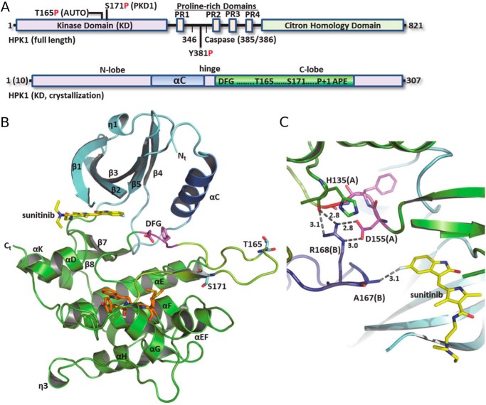
Figure 1.
Domain architecture and structure of HPK1. A, schematic of primary sequence and domains of full-length HPK1 (top) and kinase domains used for crystallization (bottom). B, subunit structure of the HPK1+0P–sunitinib KD. N-lobe, cyan ribbon; C-lobe, green ribbon; AS, yellow; and P + 1 motif, orange. C, selected interactions at the HPK1+0P dimer interface. Chain B is colored slate. Dashed lines with numbers show distance (Å).

