
Figure 3.
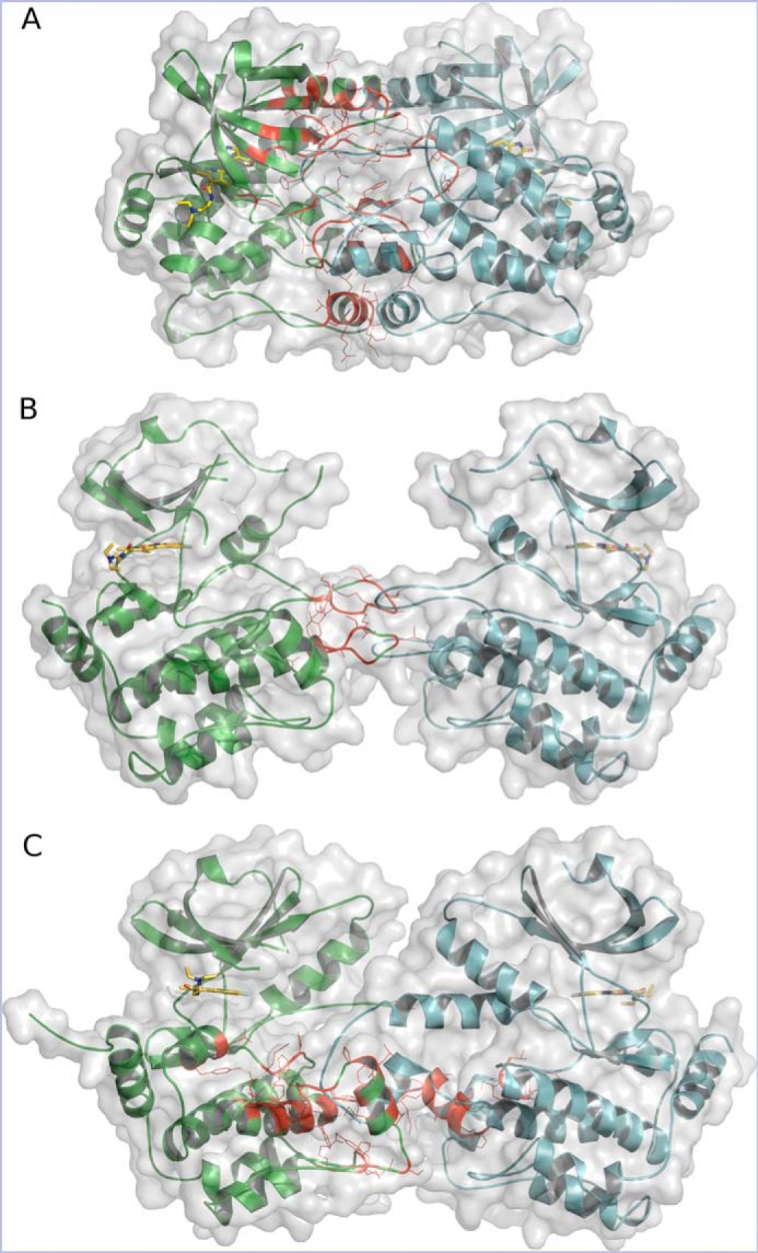
The configuration of HPK1–sunitinib dimers. A, HPK1+0P KD in a tightly closed head-to-head dimer. B, HPK1+2P KD in an open head-to-head dimer. C, HPK12PM KD in a head-to-head domain-swapped dimer. Chains A and B are colored green and cyan, respectively. Interface residues for chain A are shown as red lines, and inhibitor is drawn as yellow sticks.