Figure 1.
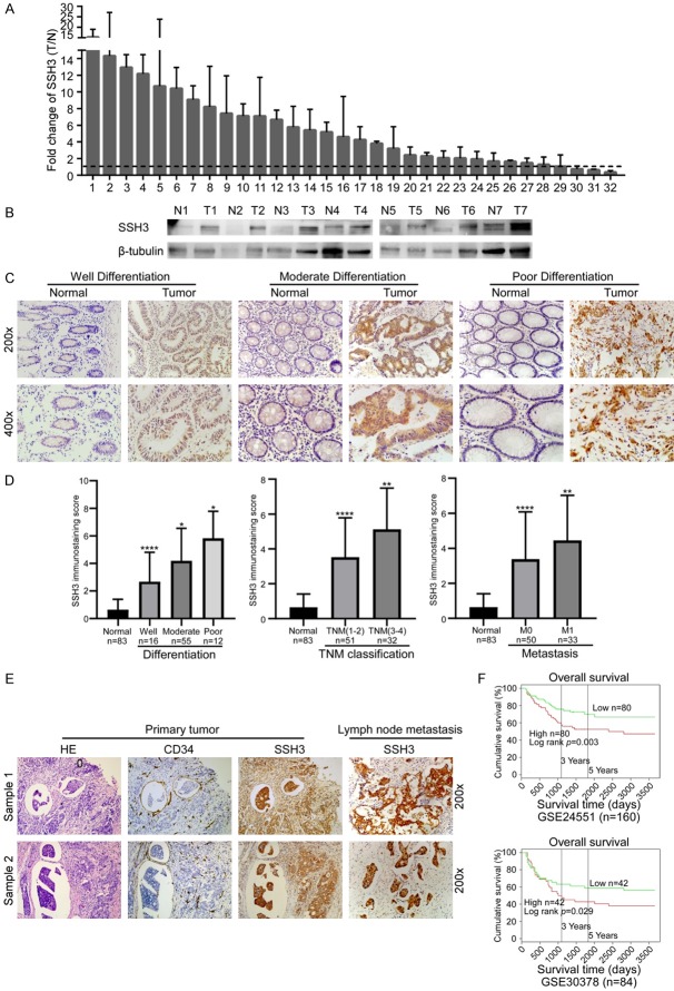
Expression of SSH3 in CRC and its correlation with CRC prognosis. A. qRT-PCR analysis of SSH3 expression in 32 paired CRC tissues; SSH3 was quantified relative to the matched adjacent no tumor tissues. Error bars represent means ± SD calculated from three parallel experiments. B. Expression analyses of SSH3 protein in 7 surgical CRC tissues and the paired normal intestine epithelial samples using Western blot. β-tubulin was used as a loading control. C. Immunostaining of SSH3 protein in CRC tissue samples and normal colorectal tissues. D. Statistical analyses of the average SSH3 immunostaining score between normal intestinal tissues and CRC specimens with different degrees of differentiation, TNM classification and Metastasis (*P < 0.05, **P < 0.01, ***P < 0.001, ****P < 0.0001). E. Immunostaining of SSH3 protein in CRC tissue samples diagnosed with tumor thrombus and the paired lymph node metastasis. F. Kaplan-Meier survival analyses published in PROG gene revealed that a higher expression of SSH3 was significantly correlated with poorer survival of patients.

