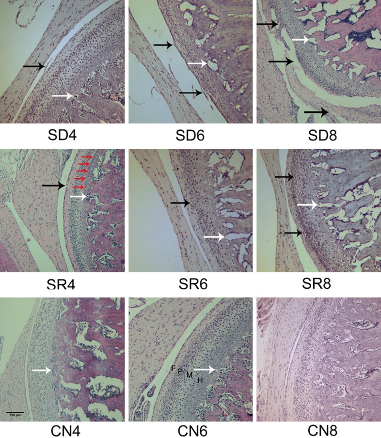
Figure 2.

Rats TMJ sections of HE staining. (A) In CN groups, the condylar cartilage displayed normal appearance, the articular surface was smooth and chondrocytes were homogenously distributed throughout the cartilage. Condylar cartilage could be clearly distinguished as four layers, fibrous layer (F), proliferating cell layer (P), mature cell layer (M), hypertrophic cell layer (H). While histopathological changes, such as loss of cartilage surface integrity reduction and disarrangement of chondrocytes, cluster formation and cell-free area (black arrows) were observed in SR and SD groups. Blood vessels at the osteochondral junction are indicated by white arrows. The tidemark was indicated by red arrows. Scale bars = 100 μm, original magnification ×100.
