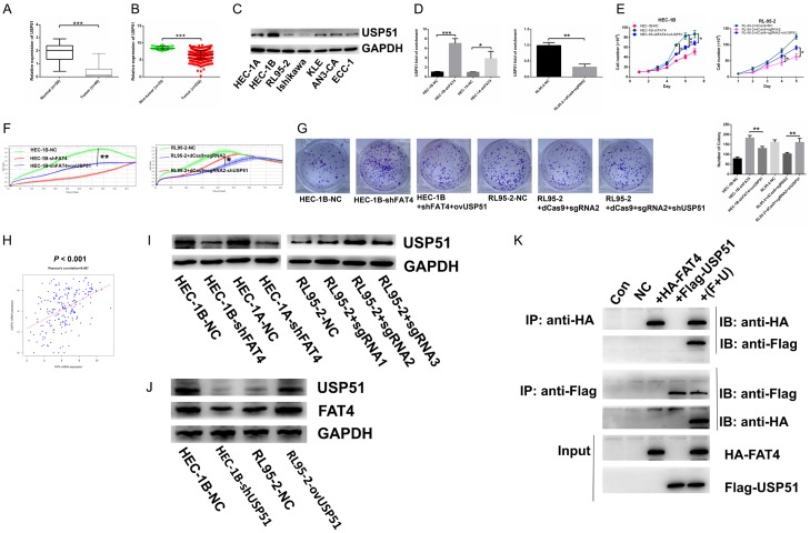
Figure 7.
Suppression of USP51 contributed to promoting tumorigenic function of FAT4 and FAT4 directly connected with USP51 to regulate mutual expression. A: mRNA expression of USP51 downregulated in EC tissues compared with normal tissues in Fudan cohort (***P<0.001, t test); B: mRNA expression of USP51 decreased in EC tissues compared with non-cancerous tissues in TCGA database (***P<0.001, t test); C: Protein expression of USP51 in 7 EC cell lines; D: To overexpress USP51 in HEC-1B-shFAT4 and HEC-1A-shFAT4 cell lines and knockdown USP51 in RL95-2+dCas9+sgRNA2 cell line (**P<0.01, ***P<0.001, t test); E-G: Increasing USP51 rescued the promoting proliferation function of silenced-FAT4 in HEC-1B cell line while HEC-1A presented no significance. Decreasing USP51 rescued the inhibiting proliferation function of upregulated-FAT4 in RL95-2 cell line (*P<0.05, **P<0.01, One-way ANOVA); H: mRNA expression of FAT4 and USP51 association based on TCGA database (Person’s correlation = 0.467); I: Validating the protein expression of USP51 in HEC-1A and HEC-1B infected with either NC or shFAT4 and RL95-2 infected with either NC or sgRNA. GAPDH was a loading control; J: Detection the protein expression of USP51 and FAT4 in HEC-1B cells transfected either with NC or shUSP51; RL95-2 cells transfected either with NC or ovUSP51 plasmid. K: CoIP assay was used to test the direct connection between FAT4 and USP51 in HEK293T cell line.

