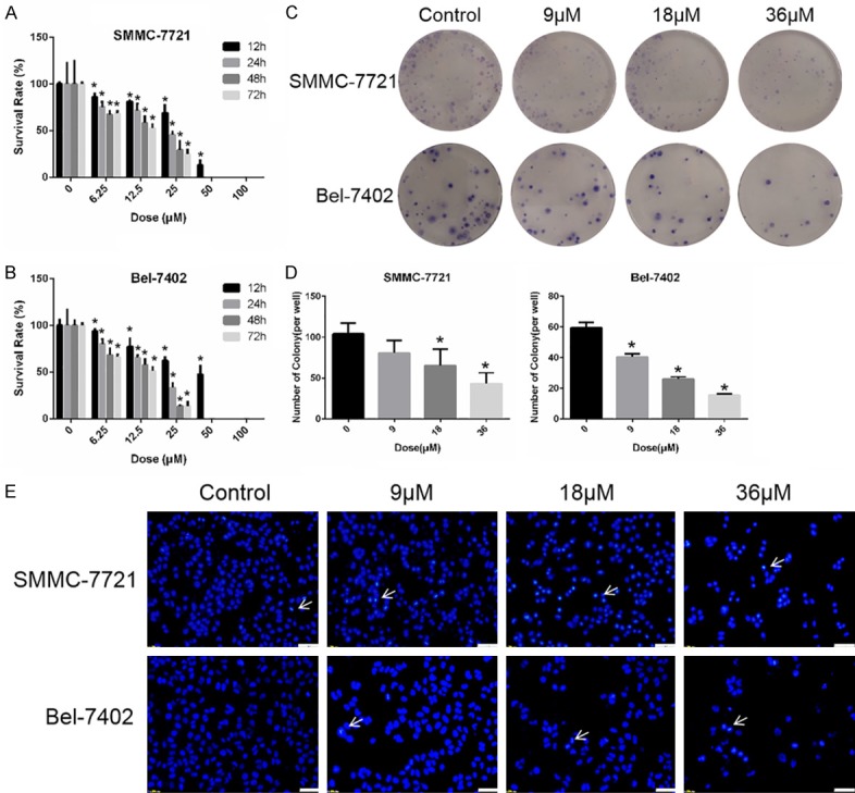
Figure 1.

Sodium demethylcantharidate effectively suppressed the proliferation of HCC Cells. (A) SMMC-7721 and (B) Bel-7402 cells were treated with various doses of sodium cantharidinate for 0, 12, 24, 48 or 72 h respectively. Cell viability was determined by SRB colorimetry. (C and D) SMMC-7721 and Bel-7402 cells were treated with sodium demethylcantharidate (0, 9, 18 or 36 µM) for 24 h. The colony formation assay was then performed to detect proliferation. (E) SMMC-7721 and Bel-7402 cells were treated with sodium demethylcantharidate (0, 9, 18 or 36 µM) for 24 h. The apoptosis of SMMC-7721 and Bel-7402 cells was detected by Hoechst staining. Bar: 50 μm. The Hoechst-positive cells (white arrows pointed) were recognized as apoptotic. The experiments were repeated at least three times. The data are expressed as the means ± SD of three experiments (*P<0.05 vs. control).
