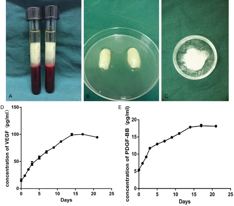
Figure 3.

PRF appearance and the release of VEGF and PDGF-BB from PRF. (A) The middle layer after centrifugation contained the PRF clot. (B) Fresh PRF isolated from blood samples. (C) Lyophilized PRF particles. ELISA results for (D) VEGF and (E) PDGF-BB released from lyophilized PRF.
