Figure 1.

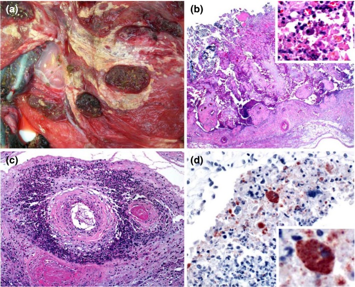
Placenta of a goat that aborted due to Chlamydia abortus infection. (a) Gross view of the chorionic side of the placenta. The membrane is thickened, with extensive multifocal to coalescing fibrinopurulent exudate. (b) Necrosis of chorioallantoic membrane, with eosinophilic cellular and karyorrhectic debris, viable and degenerate neutrophils, fewer macrophages and multifocal mineralization. Insert: higher magnification showing inflammatory cells and a trophoblast with myriad intracytoplasmic inclusions. HE. (c) Arteries within the chorioallantoic membrane showing vasculitis, thrombosis and perivascular accumulations of viable and degenerate neutrophils, macrophages, lymphocytes and plasma cells. HE. (d) Chorionic villus showing trophoblasts with myriad intracytoplasmic inclusions stained positively for Chlamydia abortus. There is also extracellular positive material. Insert: higher magnification showing positively stained trophoblasts. C. abortus immunohistochemistry.
