Figure 2.
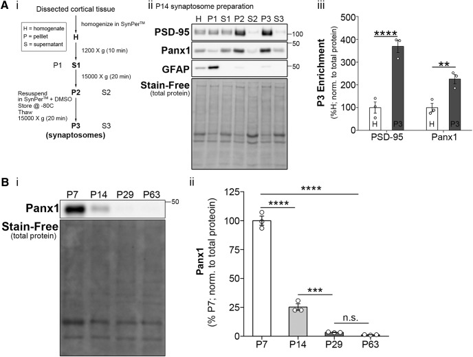
Panx1 is enriched in synaptic compartments. A, Synaptic protein extraction and isolation revealed Panx1 enrichment in cortical synaptic compartments. Ai, Protocol for synaptosome preparation from dissected cortical tissue using SynPer. Aii, Western blot of subcellular fractionations obtained from a P14 WT brain and probed with PSD-95 (top), Panx1 (second panel), and GFAP (third panel), with Stain-Free (total protein) at the bottom, demonstrating enrichment of PSD-95 in the P3 fraction (synaptosomes) and exclusion of GFAP (negative control). Aiii, Quantification of Panx1 enrichment in synaptic compartments as determined by higher immunoreactivity in P3 (synaptosomes) relative to homogenate. As expected, PSD-95 was also enriched in P3 (Panx1, p = 0.0093g6,8; PSD-95, p < 0.0001g5,7; simple-effect ANOVA with Bonferroni’s multiple-comparison test; n = 3 animals; **p < 0.01, ****p < 0.0001). B, Panx1 cortical expression is developmentally down regulated. Bi, Western blot of WT dissected whole cortical tissues from P7-P63 animals, probed with Panx1 (top), and Stain-Free (total protein) at the bottom. Bii, Panx1 expression decreased with age (age: F(3,8) = 365.9, p < 0.0001h1; n = 3 animals per group; ****p < 0.0001) with levels markedly dropping from P7 to P14 (p < 0.0001h2; P14–P29, p = 0.0006h3; P29–P63, p = 0.9604h4; one-way ANOVA with Bonferroni’s comparison test; n = 3 animals per age group; ***p < 0.001; ****p < 0.0001; n.s., not significant). Data are presented as mean ± SEM.

