Figure 1.

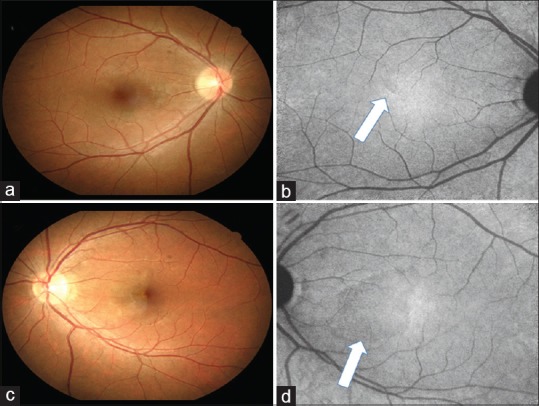
Color fundus photograph and near-infrared reflectance images of the right eye (a and b) and the left eye (c and d). Lesions (arrows) are prominent in the left eye and can be better visualized from near-infrared reflectance images

Color fundus photograph and near-infrared reflectance images of the right eye (a and b) and the left eye (c and d). Lesions (arrows) are prominent in the left eye and can be better visualized from near-infrared reflectance images