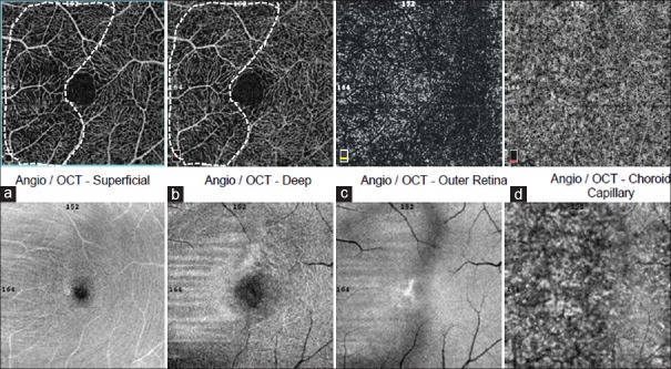
Figure 4.
Optical coherence tomography angiography and en face optical coherence tomography of the left eye. (a) Superficial capillary plexus; (b) deep retinal capillary plexus; (c) outer retina and (d) choroid capillary. Note the lesion was prominent in the left eye and similarly the vascular defects both in superficial capillary plexus and deep capillary plexus are correlated to the lesions identified from en face optical coherence tomography

