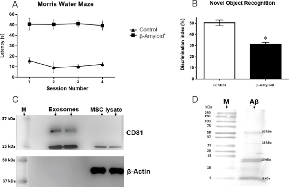
Figure 1.

Effects of Aβ1–42 administration on mouse learning and memory and exosomal marker immunodetection.
(A) Morris water maze training phase was performed four times per day for 3 days, and test session was carried out 24 hours after the last training session. Mice of the beta-amyloid group exhibited significantly shorter latency to find the platform than mice of the normal control group (no surgery). Graph data represent time to reach platform (seconds). (B) The Novel Object Recognition test shows mouse’s ability to discriminate a familiar object from a new one. Mice of the beta-amyloid group have a lower discrimination ratio than mice of the normal control group. Data graph is interaction percentage with a new object. Both Morris water maze and Novel Object Recognition tests demonstrate the model establishment. (C) Representative western blot assay for exosomes isolation verification. CD81 marker is expressed in both ultracentrifugation pellet and MSC lysate; β-actin expression was detected only in MSC lysate. Gels were run in duplicate under no reducing conditions. (D) Representative western blot assay for Aβ aggregation. Aβ1–42 peptides can aggregate in different forms with a molecular weight from 13 to 50 kDa. However, fibrillar forms did not appear. All data are expressed as the mean ± SE; *P < 0.05, vs. normal control (control) group; n = 36 for β-amyloid group and n = 8 for normal control group. MSC: Mesenchymal stem cell; M: protein molecular weight marker; s: second.
