Figure 3.

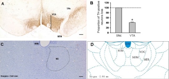
TH immunohistochemical staining of the SNc/VTA and Nissl staining of the lesion of the MD in rats.
(A) TH immunohistochemical staining images showed the comparison of dopamine neurons between the injected side (left) and non-injected side (right) in the 6-OHDA-lesioned rat. (B) Proportion of TH-immunoreactive neurons loss in SNc and VTA of the 6-OHDA-lesioned rat. (C) Nissl staining showed contrast with unlesioned side (right), damage effect of ibotenic acid injection on MD (left), and the location map of MD (D). Scale bars: 200 µm. D3V: Dorsal 3rd ventricle; MD: mediodorsal thalamic nucleus; MDM, MDC, MDL: mediodorsal thalamic nucleus, medial part, central part, lateral part; MHb: medial habenular nucleus; MTN: medial terminal nucleus; SNc: substantia nigra pars compacta; TH: tyrosine hydroxylase; VTA: ventral tegmental area.
