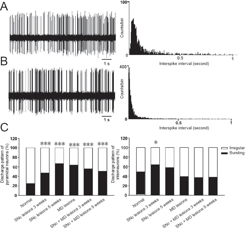
Figure 4.

Discharge patterns of neurons recorded in the mPFC of rats.
Representative sample of spontaneous activity of medial prefrontal cortex neurons showed discharge pattern of neurons and corresponding inter-spike interval histogram (bin width = 4 ms) (A, irregular discharge; B, bursting discharge). (C) Distribution of discharge pattern of mPFC pyramidal neurons or interneurons in each group. *P < 0.05, ***P ≤ 0.001, vs. normal group (one-way analysis of variance). mPFC: Medial prefrontal cortex; SNc: substantia nigra pars compacta; MD: mediodorsal thalamic nucleus.
