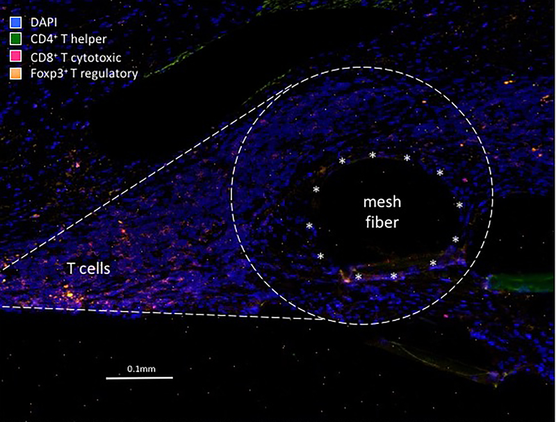
Figure 1.
Immunoflourescent micrograph demonstrating the typical T cell response to a mesh fiber in women with mesh complications. Unlike what is typically seen with the foreign body response in which macrophages immediately surround the mesh fiber (typical position marked by asterisks), T cells are observed at a distinct location away from the fiber. Image taken at 200x magnification.

