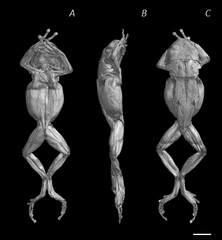
Figure 1. Reconstructed DICE µCT scan images of Phlyctimantis maculatus.

Created using N-Recon and CT-vox software. (A) Ventral, (B) lateral, and (C) dorsal view. Scale bar in white 1 cm.

Created using N-Recon and CT-vox software. (A) Ventral, (B) lateral, and (C) dorsal view. Scale bar in white 1 cm.