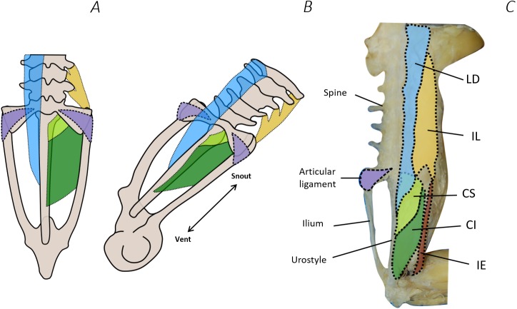
Figure 10. Comparison between Emerson’s characteristic Type IIA pelvic morphotype and traditional dissection data from Phlyctimantis maculatus.
(A) and (B) schematic diagrams adapted from Emerson (1982) and Emerson & De Jongh (1980) show dorsal and posterior-oblique dorsal views, respectively. (C) Shaded traditional dissection photograph of the dorsal spine and pelvis of P. maculatus. LD, longissimus dorsi, blue shading; IL, iliolumbaris, yellow shading; CS, coccygeosacralis, light green shading; CI, coccygeoiliacus, dark green shading; IE, iliacus externus, red shading. Articular ligament shaded purple.

