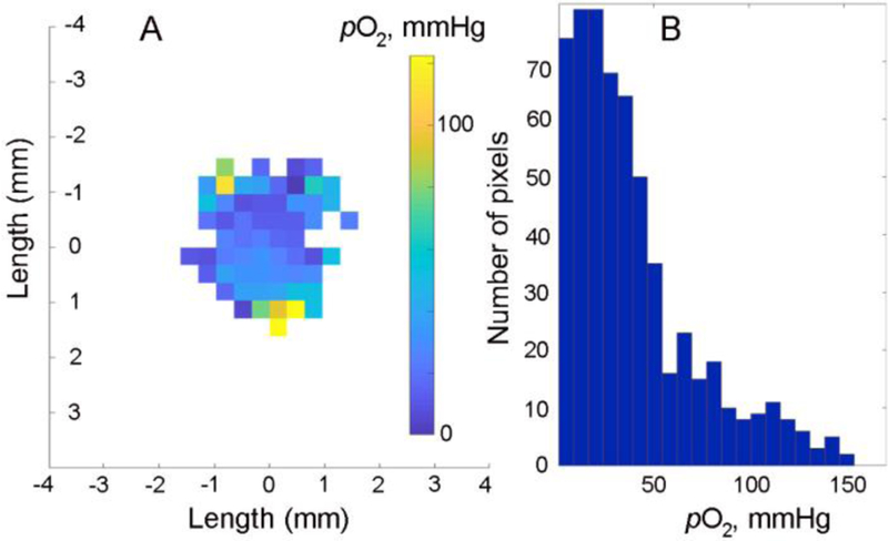
Figure 5.

In vivo 4D (1-spectral-3-spatial) rapid scan 800 MHz EPR image of oxygen distribution in a breast tumor of a PyMT tumor-bearing mouse. An image acquisition was started 25 minutes after intratissue injection of 20 µl solution of 0.75 mM probe in 10 mM phosphate buffered saline; tumor volume, 214 mm3. Data acquisition parameters were as follows: acquisition time, 16.5 minutes; number of projections, 2546; rapid scan frequency, 9.4 kHz; and maximum gradient, 3G/cm. An integral intensity threshold of 30% was implemented to remove low signal-to-noise data. A. A two-dimensional slice (xz-plane) of the image is shown. B. A histogram of pO2 distribution within the entire image.
