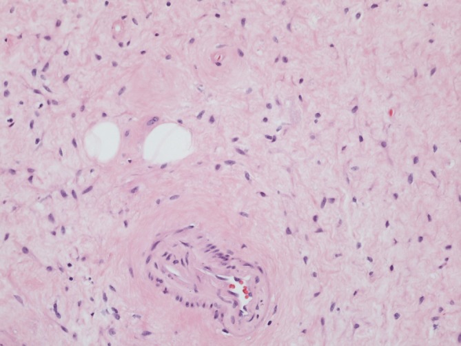
Figure 1.

Imaging (×20) of the treated tumour, hematoxylin and eosin staining. Tumour shows delicate collagenised stroma. Lower centre of the image highlights a small calibre blood vessel with subtle perivascular hyalinisation. Two entrapped adipocytes are present.
