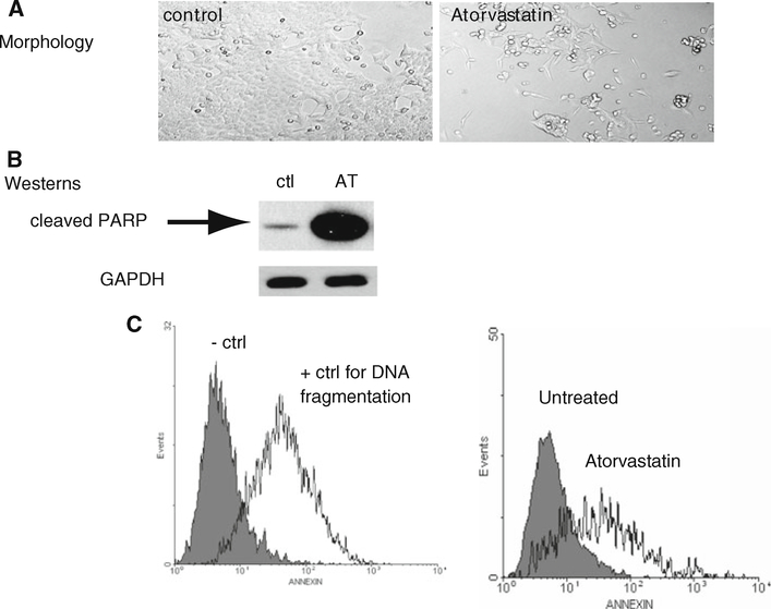
Fig. 2.
Atorvastatin increases the expression of PARP and DNA fragmentation. a Light microscopy of HCT-116 cells. Untreated (control) cells compared to cells treated with 100 μm atorvastatin for 24 h. Photomicrographs of live HCT 116 cells were taken with Leica DMIRB inverted microscope at 400× magnification. b Western-blot analysis of cleaved PARP expression in HCT-116 cells. (Ctl) untreated HCT-116 cells. (AT) HCT-116 cells treated with 100 μm atorvastatin for 24 h. c TUNEL assay with FACScan analysis of apoptosis. Representative histograms are shown. 1 × 10⁶ HCT-116 cells were treated with 100 μm atorvastatin for 24 h. TUNEL assay controls. Untreated HCT 116 cells (negative Ctl) vs. HCT-116 cells treated with (15 U) DNaseI (positive control for apoptosis). Untreated HCT 116 cells vs. HCT 116 cells treated for 24 h with 100 μm atorvastatin (n = 3 trials)

