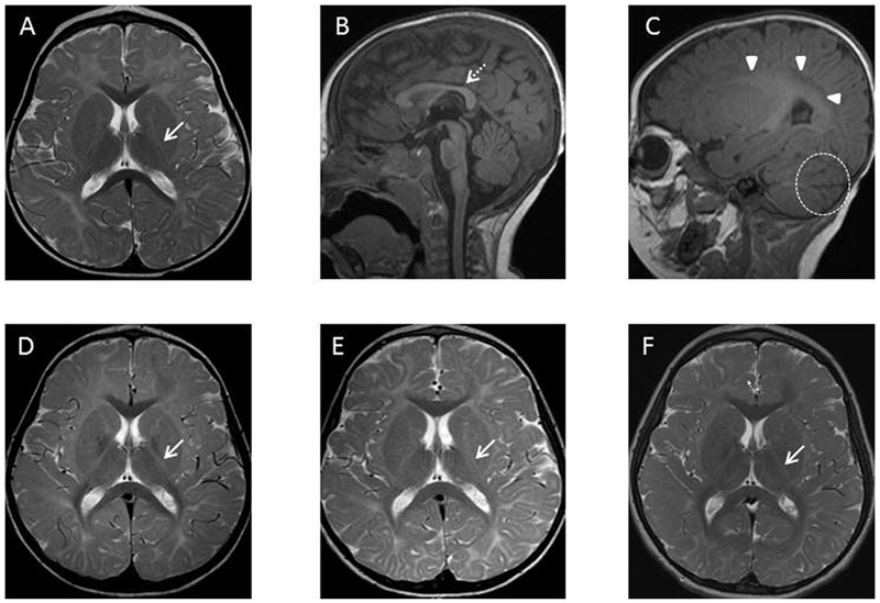
Figure 3.
Axial T2 (A, D, E, F) and sagittal T1 (B,C) images of Patient 2 at 16 months (A-C), 5 years (D), 6 years (E), and 8 years (F) of age. The initial MRI at 16 months (A-C) demonstrates delayed myelination on T1 weighted imaging (absent subcortical white matter myelination) and T2 weighted imaging (absent posterior limb of the internal capsule myelination). Follow-up imaging through 8 years of age (D-F) demonstrates no significant progress in myelination. Posterior limb of the internal capsule (A, D-F) : white arrow (solid stem). Corpus callosum (B): white arrow (dashed stem). Periventricular white matter (C): white arrowheads. Widened cerebellar fissures (C): dashed circle.

