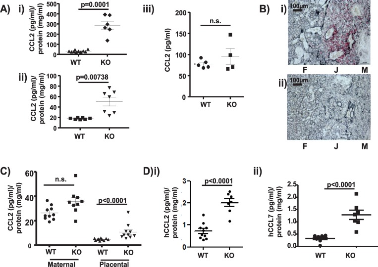
Fig 6. Placental ACKR2 limits chemokine movement from the maternal decidua to the embryo.
A) MSD-based analysis of CCL2 concentrations (normalised to total protein concentration) in plasma from i) E14.5 and ii) E15.5 WT and ACKR2–/–embryos. iii) CCL2 levels in adult pregnant mouse plasma expressed as pg/ml. Data shown here were derived from 1 of 2 repeated experiments. Unpaired Student t test without (i) and with Welch’s correction (ii) was used for statistical comparison between groups. B) In situ hybridisation showing (in red) i) ACKR2 expression (red coloration) in trophoblastic cells (grey coloration) in the E14.5 WT murine placenta and ii) lack of expression in ACKR2–/–placenta. F = foetal side of the placenta, J = foetal/maternal junction, and M = maternal decidua. Scale bar = 100 μm. C) MSD analysis of CCL2 levels (normalised to total protein concentration) in the maternal decidua and placenta of WT and ACKR2–/–E14.5 embryos. Data shown here are from 1 experiment. Similar results were independently obtained with E13.5 placenta lysates. D) Quantification of i) human CCL2 and ii) human CCL7 levels (normalised to total protein concentration) in plasma of embryos from mothers, systemically, injected i.v. with 500 ng of each chemokine. Data shown here are from 1 experiment. Unpaired Student t test without Welch’s correction was used for statistical comparison of injected human CCL2 (i) between genotypes. Data associated with this figure can be found in the supplemental data file (S2 Data). ACKR, atypical chemokine receptor; CCL, CC ligand; E, embryonic day; hCCL, human CCL; i.v., intravenously; KO, knockout; MSD, mesoscale discovery; n.s., not significant; WT, wild type.

