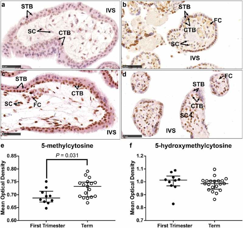
Figure 2.

Immunohistochemical labelling of 5-methylcytosine (5-mC) and 5-hydroxymethylcytosine (5-hmC) in first trimester and term tissue sections. (a) & (b). Representative images of 5-mC labelling in a first-trimester section and term tissue section, respectively. (c) & (d). Representative images of 5-hmC labelling in a first-trimester section and term tissue section, respectively. (e). Video image analysis (VIA) quantification of staining intensity revealed an increase in levels of 5-mC in tissue sections from term placenta (n = 17) compared to first trimester (n = 10). (f). There was no difference in the staining intensity of 5-hmC between first trimester (n = 10) and term tissue (n = 17) sections. Data are median and interquartile range. Significance was determined using a Mann-Whitney test. CTB: cytotrophoblast, FC: fetal capillary, IVS: intervillous space, SC: stromal cell, STB: syncytiotrophoblast.
