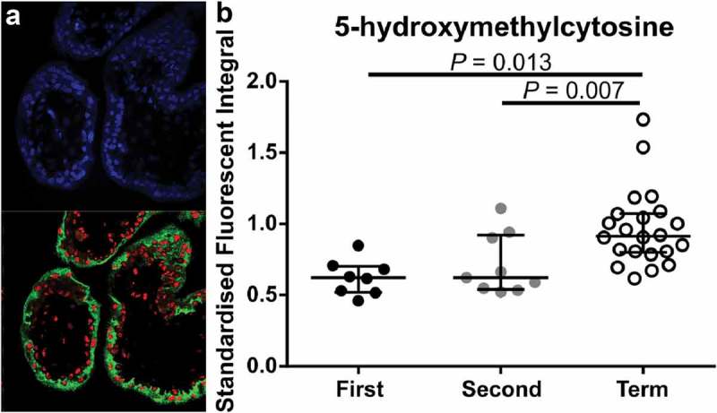
Figure 4.

Immunofluorescent labelling of 4’,6-Diamidino-2’-phenylindole (DAPI; Blue, Nuclei), 5-hydroxymethycytosine (5-hmC; Red) and Pregnancy-specific beta-1-glycoprotein 1 (PSG-1; Green, syncytiotrophoblasts (STB)). (a). Representative image of PSG-1 and 5-hmC in a first-trimester placenta tissue section. (b). Quantification of 5-hmC in STB cells across gestation using laser scanning confocal microscopy showed a significant increase in 5-hmC staining intensity in term STBs compared to first and second trimester STBs. Data are median and interquartile range. Significance was determined using an ANOVA with Tukey post-hoc comparison.
