
Figure 5.
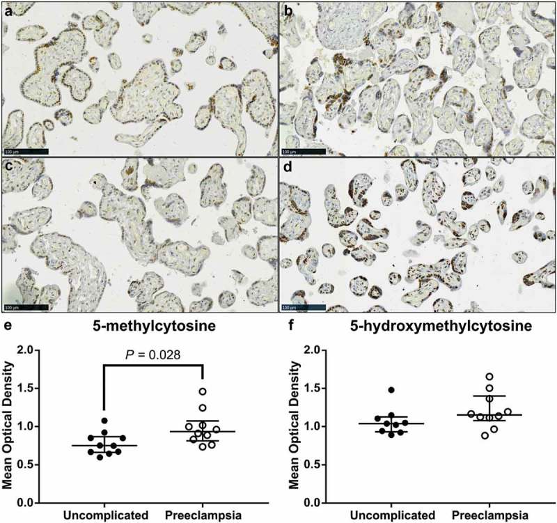
Video image analysis (VIA) of immunohistochemical staining of 5-methylcytosine (5-mC) and 5-hydroxymethylcytosine (5-hmC) in term placenta sections from pregnancies complicated by preeclampsia (PE) and uncomplicated. (a) & (c). Representative images of 5-mC staining in placenta samples from uncomplicated and PE pregnancies, respectively. (b) & (d). Representative images of 5-hmC staining in placenta in an uncomplicated and PE pregnancy, respectively. (e). Intensity of 5-mC staining was greater in tissue sections from PE pregnancies compared to uncomplicated. (f). Staining intensity of 5-hmC did not differ. Data are median and interquartile range. Significance was determined using a Mann-Whitney test.