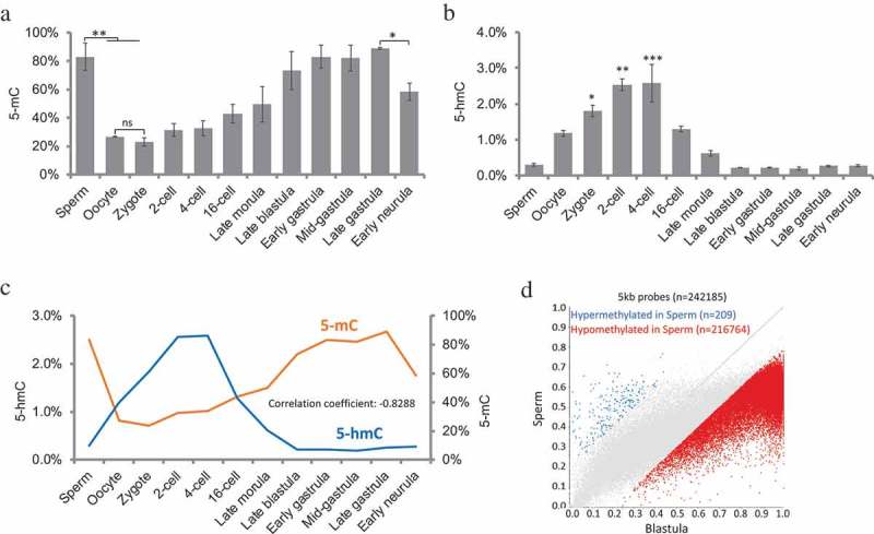
Figure 1.

Genome methylation (5-mC) and hydroxymethylation (5-hmC) in medaka during embryogenesis. (a): Constitutive global DNA methylation; one-way ANOVA analysis showed p < 0.001; two-sample t-test showed significant differences between Sperm vs. Oocyte, Sperm vs. Zygote and Late gastrula vs. Early neurula; (b): constitutive global DNA hydroxymethylation levels; one-way ANOVA analysis showed p < 0.0001, Tukey’s multiple comparisons test showed significant differences between Zygote, 2-cell, 4-cell vs. Sperm, blastula stages, and gastrula stages; (c): correlation between 5-mC and 5-hmC levels during embryogenesis. (d): differentially methylated probes between Sperm and Blastula embryos. Data represent mean ± SEM in Figure 1(a) and (b). Asterisk indicates statistical significance (*p < 0.05; **p < 0.01).
