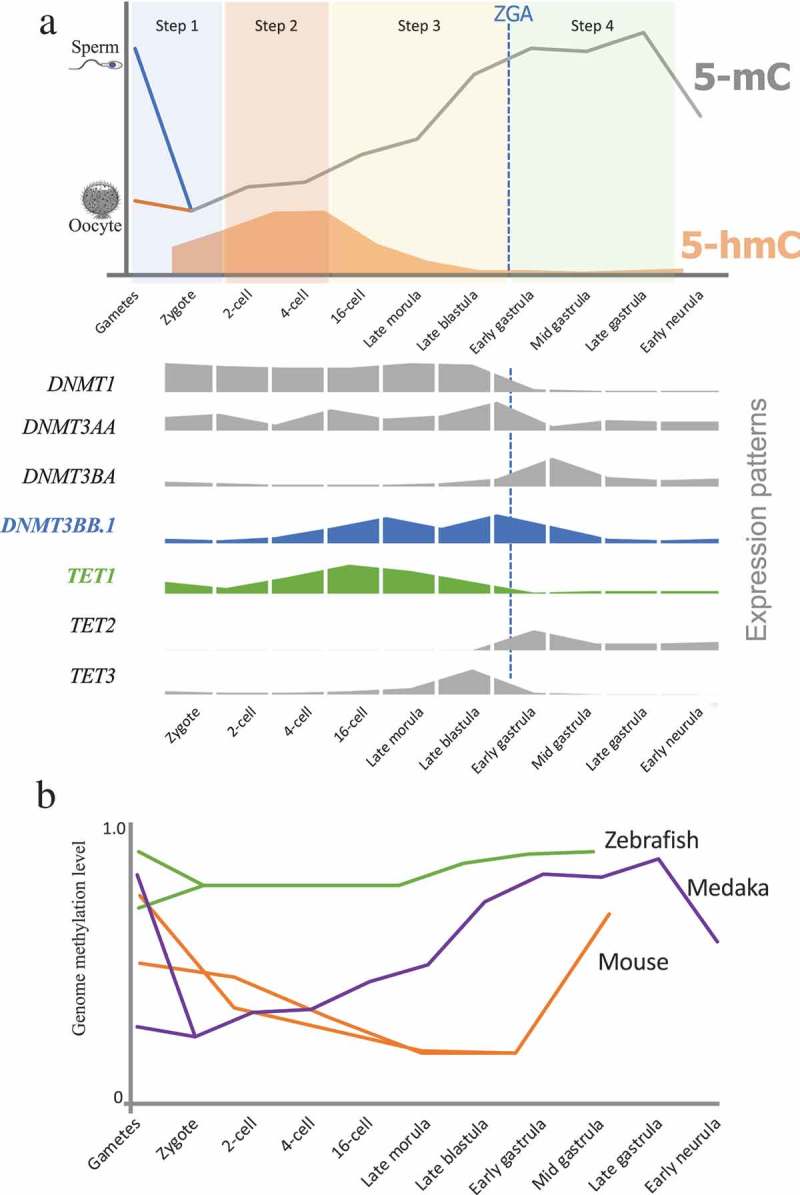
Figure 4.

DNA methylation reprogramming model during embryogenesis in medaka. (a): Sperm was hypermethylated and oocyte was hypomethylated. Paternal genomic methylation was erased in zygote (Step 1). Embryos stayed in hypomethylation status during first several cell cycles (Step 2). Global DNA methylation levels increased from 16-cell stage to Late blastula stage (Step 3). Embryos maintained hypermethylation during gastrula stages (Step 4). Global DNA methylation level decreased from gastrula to neurula stage. Relative quantification expression of genes involved in DNA methylation and hydroxymethylation were showed on lower panel. (b): A comparative epigenetic programming during embryogenesis in three model species – zebrafish [29,30], medaka, and mice [23,26].
