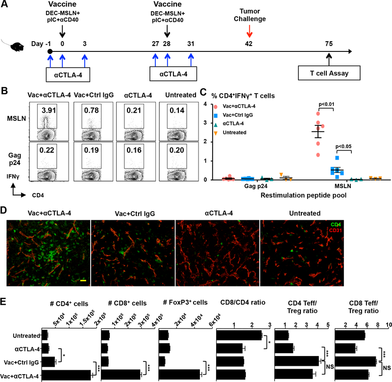
Figure 1.
Anti-CTLA–4 in combination with a protein vaccine increases intratumoral T effector cells. (A) Immunization and tumor challenge protocol. (B and C) Mice were challenged with pancreatic cancer cells (Panc02) two weeks after boost immunization. MSLN-specific CD4+ T cells were quantified in enriched TILs by intracellular IFN-γ staining ~30 days after tumor challenge. Representative FACS data (B) and quantified percentage of IFN-γ+ cells in the viable CD45+CD3+CD4+ population (C) are shown. Each dot represents tumor-infiltrating lymphocytes (TILs) pooled from 2 or 3 tumors. (D) Analysis of frozen tumor sections by immunofluorescent confocal laser microscopy. Tumors were dissected and fresh frozen in optimum cutting temperature solution (OCT), cut and stained for CD4 (green) and CD31 (red). Images were acquired with a 20× water immersion objective. Bar, 50 μm. (E) Quantification of the number of different T cell populations in TILs by flow cytometry. Data in B–E are representative of three (B and C) and two (D and E) individual experiments.

