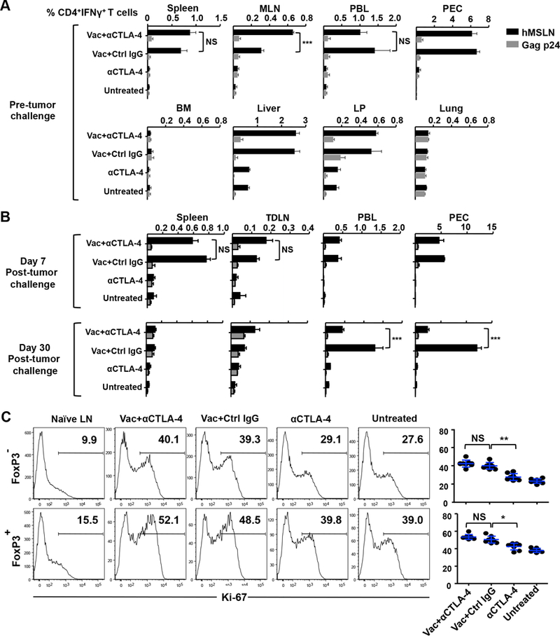
Figure 2.

Increased mobilization of vaccine-induced T effector cells from the periphery to tumor with anti–CTLA-4. (A) Two weeks after boost immunization, vaccine-induced, MSLN-specific CD4+ T cells were quantified by intracellular IFN-γ staining in lymphocytes from spleen, mesenteric lymph node (MLN), peritoneal exudate (PEC), peripheral blood (PBL), bone marrow (BM), liver, lamina propria (LP), and lungs. Percentage of IFN-γ+ cells in viable CD3+CD4+-gated cells is shown. (B) Immunized mice were challenged with Panc02 tumor cells. Seven or 30 days after tumor challenge, MSLN-specific CD4+ T cells were quantified by intracellular IFN-γ staining in spleen, tumor-draining LN (TDLN), PBL, and PEC. (C) Expression of Ki-67 in intratumoral CD4+ Teff and Treg cells. Enriched TILs were stained for CD3, CD4, and Live/Dead Aqua, followed by permeabilization, fixation, and staining for Ki-67 and FOXP3. Representative FACS data are shown. Data are representative of two or three individual experiments (n = 4 mice per group). *P < 0.01, **P < 0.01, ***P < 0.001, NS: not significant.
