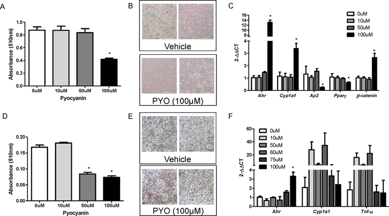
Figure 1.

Pyocyanin reduces in vitro adipocyte differentiation and induces markers of AhR activation in 3T3-L1 adipocytes. (A) Oil Red O (ORO) absorbance (510 nm, day 8 of differentiation protocol) of 3T3-L1 adipocytes treated with vehicle (0 µM) or pyocyanin. (B) Representative images of cells (day 8 from (A)) incubated with vehicle or pyocyanin (PYO). (C) mRNA abundance of AhR, CYP1a1, aP2, PPARγ, or β-catenin in 3T3-L1 adipocytes incubated with vehicle (0 µM) or pyocyanin during the differentiation protocol. (D) ORO absorbance of differentiated (day 8) 3T3-L1 adipocytes incubated with vehicle (0 µM) or pyocyanin for 24 hours. (E) Representative images of differentiated 3T3-L1 adipocytes from (D) incubated with vehicle or pyocyanin. (F) mRNA abundance of AhR, CYP1a1, or TNF-α mRNA abundance in differentiated 3T3-L1 adipocytes incubated with vehicle (0 µM) or pyocyanin for 24 hours. Scale bar in (B) and (E) is 200 μm. Data are mean ± SEM from n = 3–8 wells/treatment. *, P<0.05 compared to 0 μM (VEH).
