Figure 3.
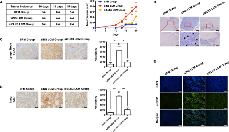
The suppression of ELK3 in LECs diminishes the ability of LECs to promote tumor growth in vivo. Athymic nude mice (female, 5–6 weeks of age, 18–20 g) were pretreated with LCM harvested from siNS- or siELK3-transfected LECs or SFM subcutaneously for 2 weeks, as described in the Material and Methods. Then, 2 × 10⁶ MBA-MB-231 cells were mixed with 3.7 × 10⁶ siNS- or siELK3-transfected LECs and Matrigel and inoculated into the mammary fat pads of the corresponding nude mice. MDA-MB-231 cells with siNS LECs were inoculated into siNS LCM-pretreated mice (siNS LCM group), and MDA-MB-231 cells with siELK3 LECs were inoculated into siELK3 LCM-pretreated mice (siELK3 LCM group). For the control, 2 × 10⁶ MDA-MB-231 cells were inoculated into SFM-pretreated mice (SFM group). (A) Tumor incidence was examined in four mice from each group on the indicated days after inoculation (Left). Tumor size was measured at the indicated date after inoculation and presented as a graph (Right). (B) Matrigel plugs excised from mice 20 days after inoculation were fixed and stained with H&E. Scale bar represents 50 μm. LNs (C) or lungs (D) were isolated from mice from each group, and immunohistochemical staining was performed to detect human cytokeratin-7 (CK7). Scale bar represents 50 μm. (E) LNs from each group were analyzed for the expression of mouse CD31 by immunofluorescence staining. Scale bar represents 50 μm. Error bars represent the standard error from three independent experiments, and each experiment was performed using triplicate samples. *P < 0.05, **P < 0.01 ***P < 0.001 and ****P < 0.0001 (Student’s t-test).

