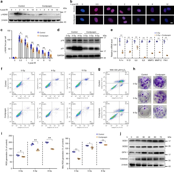
Fig. 2.
Cordycepin decreases cell senescence and SASP in vitro. a Western blot analysis of γ-H2AX expression in fibroblasts after 5 Gy radiation. b Representative images of nuclei γ-H2AX in fibroblasts 0–12 h after 5 Gy radiation. This test was repeated three times. Representative images were shown. c Average number of γ-H2AX per nucleus 0–12 h after 5 Gy radiation (n = 7). d Western blot analysis of p16 and p21 levels in irradiated control or cordycepin-treated fibroblasts 7 days after radiation. This test was repeated three times. Representative images were shown. e Quantification of mRNA expression for senescence secretory phenotype in irradiated control or cordycepin-treated fibroblasts 7 days after radiation (n = 3). f Fibroblasts were treated with cordycepin for 72 h and harvested for detection of apoptosis using flow cytometry 24 h following exposure to radiation. g Fibroblasts were treated with cordycepin for 72 h and harvested for detection of apoptosis using flow cytometry 12 h following H2O2 treatment. h Representative images of fibroblasts colonies generated in survival assays following 0/5/8 Gy radiation. i Analysis of reactive oxygen species (ROS) levels and mitochondrial superoxide by H2DCF-DA or MitoSOX Red staining 24 h after radiation in fibroblasts pretreated or not (control) with cordycepin (n = 3). j SOD1, SOD2, GPX-1, and Catalase expression levels in whole cell lysates from cordycepin-treated fibroblasts for the indicated times. Bars represent 10 μm (b). Data in c, e, and i represent the means ± S.D. (*P < 0.05, **P < 0.01; student’s t-test)

