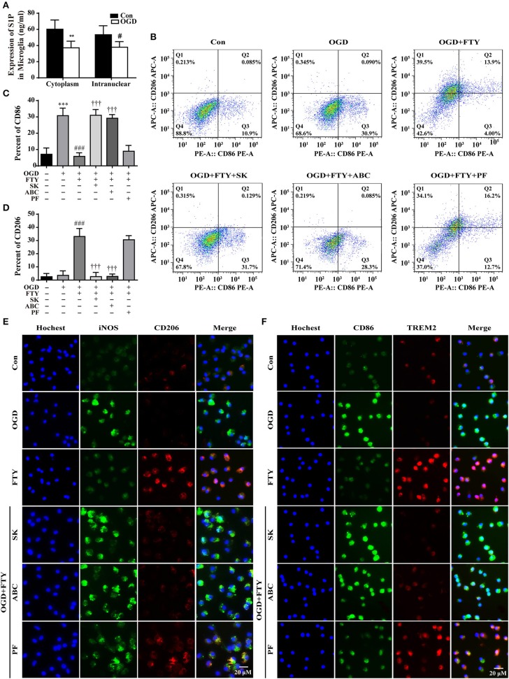
Figure 4.
Intra-nuclear SphK2-mediated phosphorylation is necessary for FTY720-facilitated the switch of microglial polarization from M1 to M2. (A) The expression of S1P was analyzed by ELISA. n ≥ 4 per group, **p < 0.01, compared to the Con group in Cytoplasm; #p < 0.05, compared to the Con group in intra-nuclear, by Student's t-test with unpaired. Fluorescence-activated cell sorting analysis of the microglia in the OGD, OGD+FTY720, OGD+FTY720+SK, OGD+FTY720+ABC, and OGD+FTY720+PF groups. Surface expression of CD86 and CD206 was detected in microglia by flow cytometry. The percentage of CD86 (B,C) and CD206 (B,D) in the microglia was analyzed. n ≥ 4 per group, ***p < 0.001, compared to the Con group; ###p < 0.001, compared to the OGD group; †††p < 0.001, compared to the OGD+FTY group, by one-way ANOVA and Tukey's test. SK and ABC reversed FTY720-induced changes in the protein expression of the M1 markers (iNOS and CD86, green) and the M2 markers (CD206 and TREM2, red) by immunofluorescent staining (E,F). PF failed to affect FTY720-induced changes in the protein expression of the M1 markers (iNOS and CD86, green) and the M2 markers (CD206 and TREM2, red) (E,F). Scale bar = 20 μm. All data are presented as means ± SEM.

