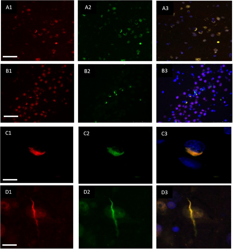
FIGURE 2.
Co-localisation of TDP-43 and hnRNP E2. Double-labeling immunofluorescence shows inclusions positive for TDP-43 (red) (A1) and hnRNP E2 (green) (A2) in the frontal cortex of a subtype A case, the merged image (A3) shows numerous areas of co-localisation (case ID BBN_15298). Co-localisation is also seen in the hippocampus, panels (B1–B3) show a subtype A case (case ID 482 BBN_4568). Higher magnification images demonstrate the complete co-localisation of the TDP-43 and hnRNP E2 in a perinuclear inclusion from the subtype A case (C1–C3) and along a dystrophic neurite in a subtype C case (D1–D3) (case ID BBN_15303). Scale bar represents 100 μm in panels (A1–A3,B1–B3), 25 μm in panels (C1–C3,D1–D3).

