FIGURE 1.
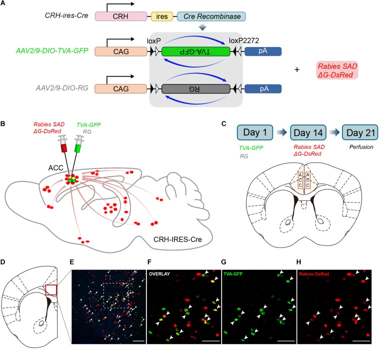
Monosynaptic inputs to the ACC CRH neurons using the rabies virus and CRH-ires-Cre knock-in mice. (A) The AAV helper virus and EnvA pseudotyped glycoprotein (G)-deleted rabies virus. (B) Combination of the two virus system and CRH-ires-Cre mice allows for brain-wide labeling of monosynaptic inputs (red) to CRH neurons in the ACC. (C) Timeline of virus injection for retrograde trans-synaptic tracing and the schematic of the anatomical localization of the ACC. (D–H) Representative confocal images of the injection site. As the starter cells express both GFP and DsRed fluorescent proteins, they are shown in yellow in the image. Panel (D) shows the diagram of the ACC section. Red box indicates the region shown in (E). Panels (F–H) show enlarged views of the white-boxed region in (E). Scale bars: 100 μm in (E) and 50 μm in (F–H).

