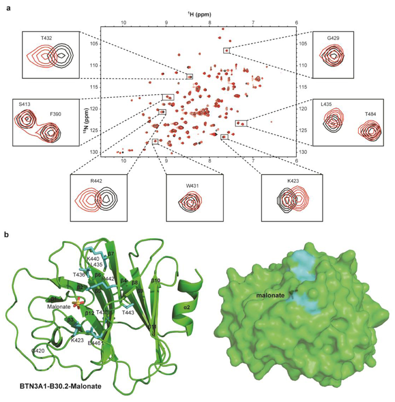
Figure 5. NMR chemical shift perturbation analysis of BTN3A1-B30.2 domain with malonate.
a) Overlay of 1H 15N HSQC spectra of 15N-labelled 0.3mM BTN3A1-B30.2 domain in the absence (black) and presence of 100mM sodium malonate (red). Black boxes highlight a close-up view of peaks corresponding to BTN3A1-B30.2 residues that undergo chemical shifts upon HMBPP/cHDMAPP binding. b) Surface mapping of perturbed residues of B30.2 (shown as stick format) upon malonate binding. Residues are coloured based on small (cyan), medium (orange) and large (red) chemical shift perturbations. Malonate is shown as stick format.

