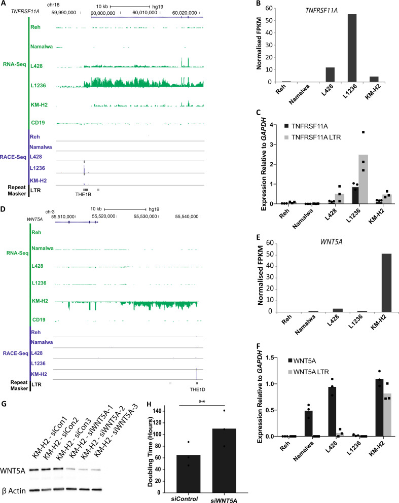
Fig. 3.
TNFRSF11A and WNT5A are expressed from active THE1B and THE1D LTRs. a UCSC genome browser screenshot showing a THE1B LTR acting as a promoter for the TNFRSF11A gene in L1236 and L428 cHL cell lines but not in primary CD19+ B cells [27]. b Normalised RNA-Seq FPKM values showing expression of TNFRSF11A. c qPCR gene expression analysis showing expression of a transcript between exon 2 and 3 and also between the upstream LTR and exon 2. Three biological replicates (p < 0.05 L1236 vs. control cell lines, paired Student's t-test). d UCSC genome browser screenshot showing an LTR identified by RACE-Seq in the KM-H2 cell line which produces a transcript of the WNT5A gene, shown by RNA-Seq read alignment. Compared to Primary CD19+ B cell RNA-Seq [27]. e Normalised RNA-Seq FPKM values showing expression of WNT5A. f qPCR gene expression analysis showing expression of a transcript between exon 2 and 3 and also between the upstream LTR and exon 2 of WNT5A. Three biological replicates (p < 0.01 KM-H2 vs. control cell lines, paired Student's t-test). g WNT5A protein measured by western blot following siRNA knockdown compared to non-targeting control. h Cell doubling time following siRNA knockdown of WNT5A. Three biological replicates (p < 0.01, paired Student's t-test)

