FIGURE 3.
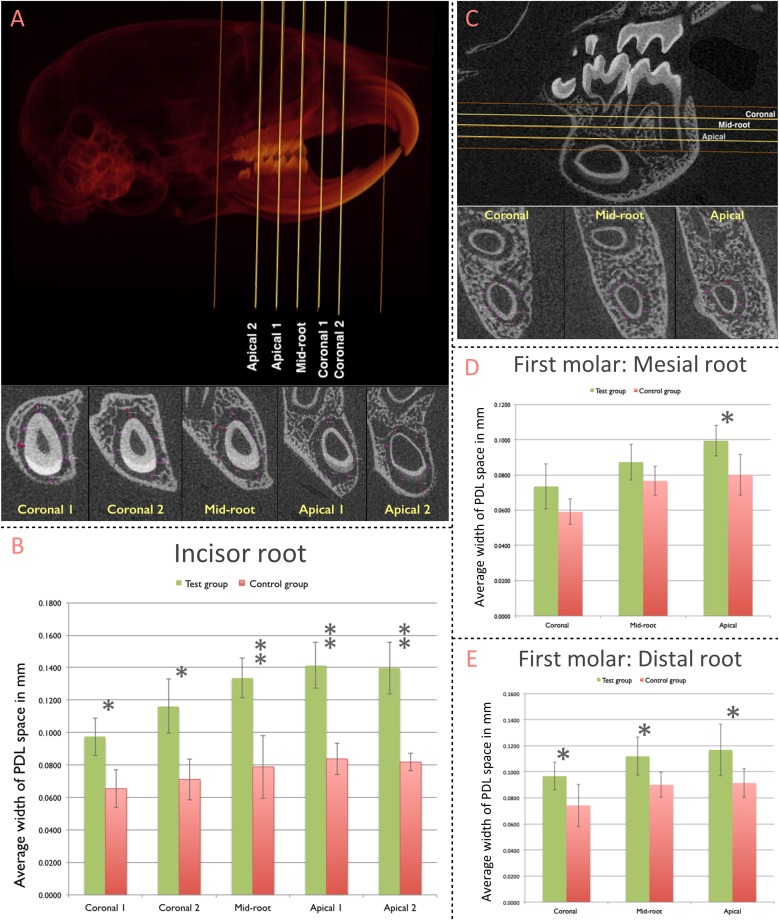
Contribution of pnPRX1+ cells to the post-natal development of the molar and incisor periodontium. (A) The width of PDL space around the root of the right mandibular incisor was measured in five transverse slices (Coronal 1, Coronal 2, Mid-root, Apical 1, and Apical 2 slices; top image). These transverse slices were generated based on the two indicated reference points (orange lines): the most coronal level of the alveolar bone (coronal reference) and the most apical point of the tooth socket (apical reference). The linear measurements of the width of PDL space were performed at 8 different points within each section (bottom image, purple arrows). The measurements at the enamel surface (buccal measurement in the most of slices) were excluded from the analysis. All 8 linear measurements were averaged and such average was considered as the width of the PDL space for each section. (B) Comparison of the width of PDL space of the mandibular incisor tooth between the test and the control groups. (C) The width of the PDL space around mesial root of right mandibular first molars was measured in three transverse slices (Coronal, Mid-root, and Apical - yellow lines) generated based on two reference points (orange lines): the furcation entrance of the tooth (coronal reference) and the apex of the mesial root (apical reference). Similar methodology was used to select the slices for the distal root of the first molars. The linear measurement was performed at 8 different points, similarly, to what was performed for the incisor tooth. (D) Comparison of the width of PDL space of mesial root of the mandibular first molar; and (E) distal root of the mandibular first molar between test and control groups. (∗p < 0.05, ∗∗p < 0.001).

