FIGURE 4.
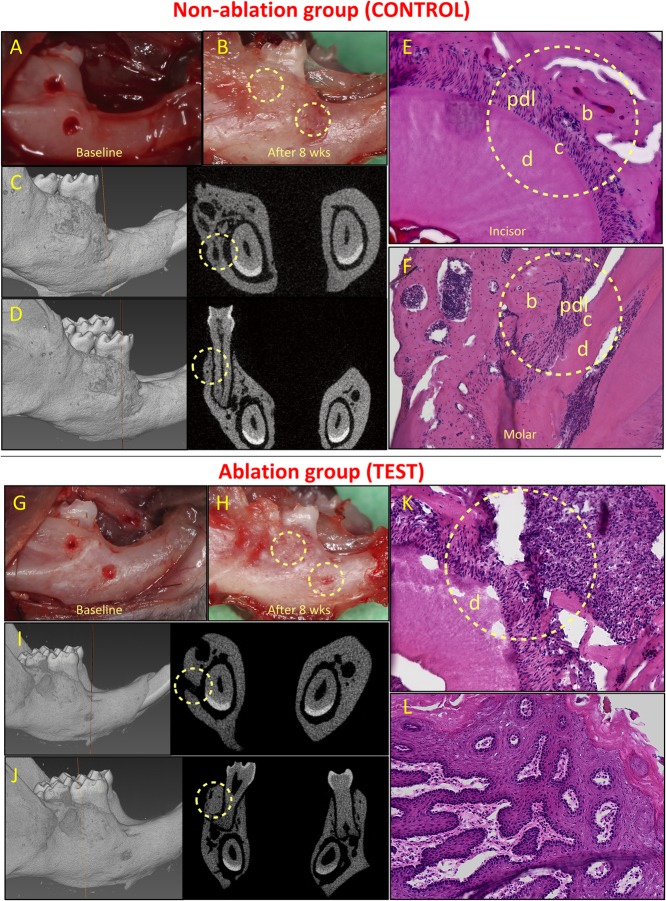
Healing of non-critical defects in non-ablation (images above the red line; A–F) and ablation (images below the red line; G–L) groups (n = 5). Two defects were created around the mandibular incisor and first molar teeth in both groups (A and G). In the non-ablation group, healing of both defects was observed clinically for all animals (B). Micro-CT analysis confirmed the healing of incisor (C) and molar (D) defects, and histological analysis confirmed regeneration of the periodontal incisor (E) and molar (F) defects. In the ablation group, lack of healing was observed for incisor defects in all animals (H). Micro-CT (I) and histological (K) analyses confirmed lack of periodontal regeneration for incisor defects in this group. The molar defects were mainly healed by excessive and irregular bone formation (H, J, L). b, bone; c, cementum; d, dentine; and pdl, periodontal ligament.

