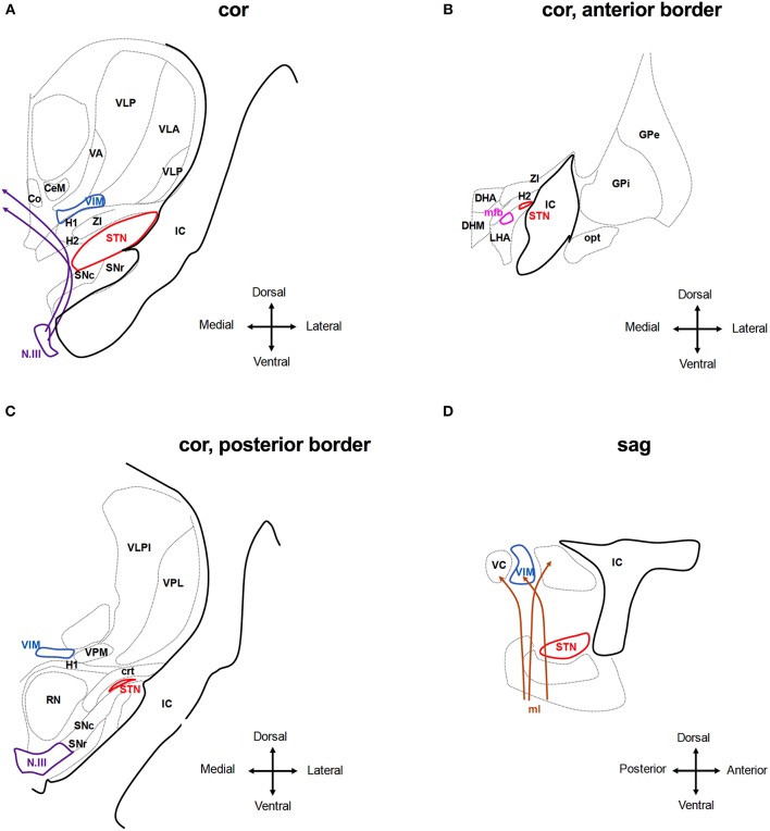
Figure 3.
Anatomical relationship of the subthalamic nucleus (STN) and the ventral intermedius nucleus (VIM) to adjacent structures. The schematic shows coronar (A–C) and sagittal (D) planes through the basal ganglia at the level of the STN and VIM. Co, Commissural nucleus; CeM, central medial thalamic nucleus; VA, ventroanterior thalamic nucleus; VC, ventrocaudal nucleus; VLP, ventrolateral posterior thalamic nucleus; VPM, ventroposterior medial thalamic nucleus; IC, internal capsule; SNr, Substantia nigra pars reticulate; and SNc compacta; H1, H2, H1 and H2 Fields of Forel; ZI, zona incerta; N.III, nucleus of the third cranial nerve; DHA, dorsal hypothalamic area; DHM, dorsomedial hypothalamic nucleus; LHA, lateral hypothalamic area; mfb, medial forebrain bundle; opt, optic tracts; RN, red nucleus; crt, cerebello-rubro-thalamic tract. Stimulating the tissue medial and dorsal to the STN activates the H1 and H2 fields of Forel and the ZI and may reach to the medio-dorsal thalamic nuclei incl. the Co, CeM, and VIM. Deflection of the field to more ventral areas will activate the fibers of the N.III and the SN (A). Anterior of the STN, stimulation may activate hypothalamic nuclei and mfb as well as the IC (B,D). At the posterior border of the STN, stimulation may activate the RN and ml, in particular, if the tissue medial of the STN is activated. Stimulation of tissue dorsal of the STN may activate the crt. (C,D). Adjusted from Mai et al. (55).

