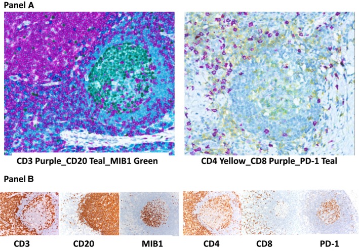
Figure 2.

Panel A, Multiplex immunohistochemistry. Three stains can simultaneously detect different proteins in formalin‐fixed, paraffin‐embedded sections of a reactive lymph node. Left: Expression of CD3 (purple) in T cells, CD20 (teal) in B cells, and both CD20 and MIB1 (green) in a large fraction of germinal centre B cells, in different subcellular locations (CD20 in the membrane and MIB1 in the nucleus). Right: Expression of CD4 (yellow) in helper T cells, CD8 (purple) in cytotoxic T cells, and both CD4 (yellow) and programmed cell death 1 (PD‐1) (teal) in a large fraction of germinal centre T cells (merging into green). Panel B, Standard immunohistochemistry. Different tissue sections of a lymph node are stained with CD3, CD20, MIB1 and CD4, CD8, PD‐1. Left: Expression of CD3 (diffuse in the paracortical area and scattered in the germinal centre), CD20 (diffuse in the follicle mantle and scattered in the germinal centre), and MIB1 (restricted to germinal centre cells). Right: Expression of CD4 and CD8 (diffuse in the paracortical area). CD4‐positive cells are present in the germinal centre. A fraction of germinal centre cells also express PD1. Images were acquired with the Aperio ScanScope XT Virtual microscopy system and ImageScope Slide Viewing software (Leica Biosystems)
