. 2019 Mar 13;18:154–164. doi: 10.17179/excli2018-1928

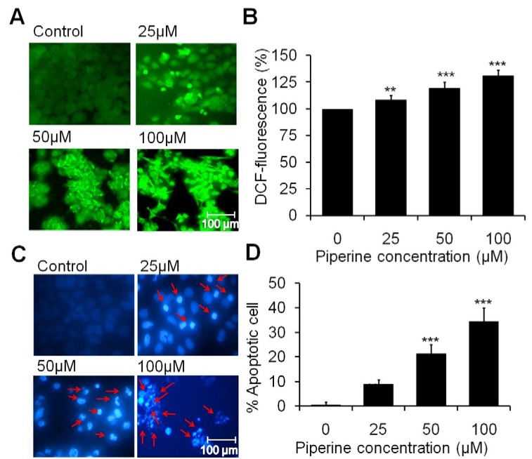
Figure 2. Piperine induces intracellular ROS generation and nuclear condensation in HeLa cells. (A) The photomicrographs show induction in intracellular ROS on HeLa cells at 25 μM, 50 μM and 100 μM concentrations of piperine. (B) Quantitative data represents the percentage of fluorescence intensity on HeLa cells with respect to the control. (C) Photomicrographs exhibits the characteristic fragmented and condensed nuclei (indicated by arrow) in piperine treated HeLa cells. (D) Statistical data expressed as percent of apoptotic cells as compared to the control. Minimum three independent experiments were performed and the values are expressed as means ± SEM, **p < 0.01 and ***p < 0.001 as compared to the control.

Figure 2