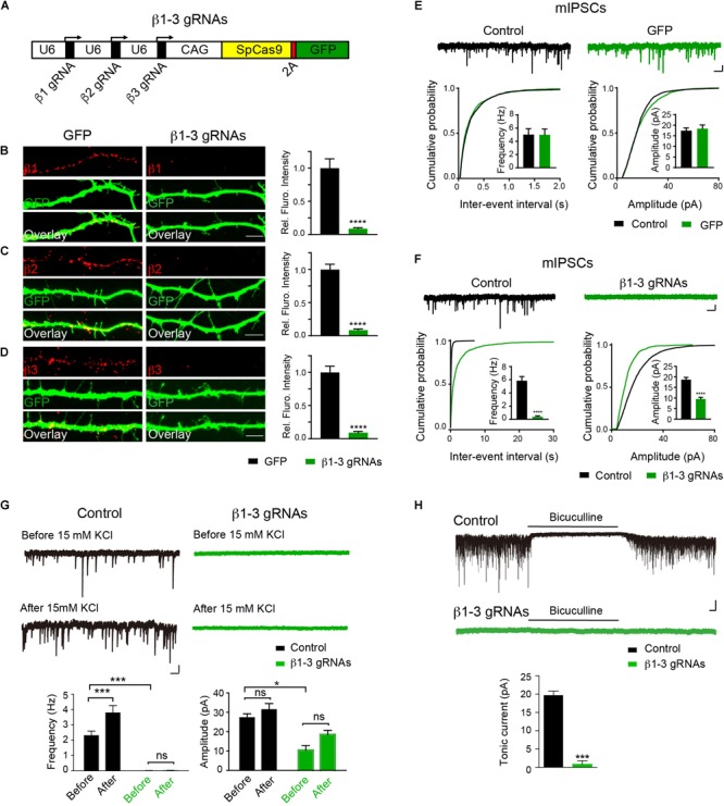
FIGURE 1.

Single-cell KO of the GABAAR β1-3 subunits eliminated GABAergic synaptic transmission. (A) Schematic of β1-3 gRNA vector. (B–D) Representative images showed the loss of β1 (B), β2 (C), or β3 (D) subunits in hippocampal neurons expressing β1-3 gRNA vector as compared to neurons expressing the empty gRNA vector (GFP) (GFP, n = 15; β1-3 gRNAs, n = 15; ∗∗∗∗p < 0.0001, t-test; N = 3). Scale bar, 5 μm. (E) Expression of the empty gRNA vector (GFP) in hippocampal neurons did not change inhibitory synaptic transmission (control, n = 10; GFP, n = 9; N = 3; t-test; p > 0.05 for mIPSC frequency and amplitude; Kolmogorov-Smirnov test was used for cumulative graphs, p > 0.05 for both conditions). Scale bar, 500 ms, 20 pA. (F) Expression of β1-3 gRNA vector in hippocampal neurons essentially eliminated inhibitory synaptic transmission (control, n = 19; β1-3 gRNAs, n = 21; N = 5; t-test, ∗∗∗∗p < 0.0001 for both frequency and amplitude; Kolmogorov-Smirnov test was used for cumulative graphs, ∗∗∗∗p < 0.0001 for both conditions). Scale bar, 500 ms, 20 pA. (G) 15 mM KCl significantly increased the mIPSC frequency in control neurons but not in neurons expressing β1-3 gRNAs at DIV 12-15 (control, ∗∗∗n = 16; p < 0.001 for frequency, p > 0.05 for amplitude; β1-3 gRNAs, p > 0.05 for frequency, p > 0.05 for amplitude; n = 19; For β1-3 gRNA amplitude analysis, only 5 out of 19 cells had mIPSC events for analysis; N = 2; one-way ANOVA followed by the Bonferroni test). Scale bar, 500 ms, 20 pA. (H) GABAAR-mediated tonic currents in control and β1-3 gRNAs-expressing neurons at DIV 14-17 (Control, n = 23; β1-3 gRNAs, n = 15; N = 3; ∗∗∗p < 0.001, t-test). Scale bar, 10 s, 50 pA. n represents the number of cells analyzed and N represents the number of independent experiments.
