
FIGURE 4.
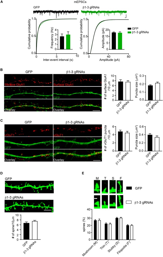
Loss of GABAergic transmission in individual neurons did not change glutamatergic transmission. (A) mEPSCs recording showed loss of GABAARs in individual neurons did not change glutamatergic transmission. Hippocampal neurons were transfected with β1-3 gRNAs at DIV3 and recorded at DIV14-17 (n = 25 for GFP and β1-3 gRNAs; p > 0.05 for mEPSC frequency and amplitude, t-test; Kolmogorov-Smirnov test was used for cumulative graphs, p > 0.05 for both conditions, N = 4). Scale bar, 100 ms, 20 pA. (B) Single-cell genetic deletion of GABAARs did not change the expression levels of surface GluA1 (GFP, n = 17; β1-3 gRNAs, n = 19; p > 0.05 for both conditions, t-test; N = 3). GFP was not immunolabeled with anti-GFP antibodies. Scale bar, 5 μm. (C) Single-cell genetic deletion of GABAARs did not change the vGluT1 puncta (GFP, n = 24; β1-3 gRNAs, n = 28; p > 0.05 for both conditions, t-test; N = 3). GFP was not immunolabeled with anti-GFP antibodies. Scale bar, 5 μm. (D) The density of dendritic spines was not altered in hippocampal neuron expressing β1-3 gRNAs at DIV 18 (GFP, n = 26; β1-3 gRNAs, n = 35; p > 0.05, t-test; N = 3). GFP was immunolabeled with anti-GFP antibodies to boost the fluorescence (green). Scale bar, 5 μm. (E) Normal spine types in hippocampal neurons expressing β1-3 gRNAs (GFP, n = 26; β1-3 gRNAs, n = 28; p > 0.05, t-test; N = 3). GFP was immunolabeled with anti-GFP antibodies to boost the fluorescence (green). Scale bar 1 μm. n represents the number of cells analyzed and N represents the number of independent experiments.