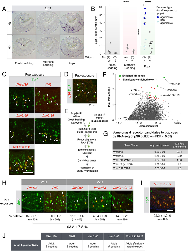
Figure 2: Pup cues activate seven VRs in the adult VNO.
(A,B) Egr1 induction in the VNO of virgin male and female mice upon pup exposure. The number of Egr1+ VNO neurons identified in these experiments is shown in (B). Each data point represents one animal. Number of animals used is indicated in the graph. ***p < 0.001 by t-Test. Dark lines indicate the mean, and light lines indicate the quartiles.
(C) Top four panels: double FISH with RNA probes to Egr1 and candidate VRs led to the identification of five VRs responding to pup cues. Arrows indicate cells in which Egr1 (green) and VR signals (red) co-localize. Bottom panel: double FISH with RNA probes to Egr1 (green) and a mix of the five VR probes shown in panels above. White arrows point to co-labeled neurons, and green arrows indicate Egr1+ neurons that express none of the 5 identified VRs.
(D) Immunofluorescence with anti-pS6 antibody and in situ hybridization with RNA probe to Egr1 demonstrates co-expression (arrows) in VNO neurons activated by pup cues (94.7 ± 2.2 % of Egr1+ neurons are pS6+; mean ± SEM; 699 Egr1+ cells from 3 animals examined).
(E) Experimental pipeline for immuno-purification of phospho-ribosome associated RNAs from VNOs of animals exposed to pup cues.
(F) MA (log2 difference over signal strength) plot showing the enrichment of specific VR in the VNOs of virgin males exposed to pup stimuli compared to fresh bedding. Red dots represent genes significantly (p < 0.1) over-represented in pup-exposed VNO samples. Green dots represent enriched VR genes.
(G) List of VRs identified in the pS6-based screen. The screen failed to identify the sparsely represented V1ri9 identified by the candidate VR screen (shown in C).
(H) Comprehensive set of VRs activated by pup cues. Double FISH with individual VR and Egr1 probes. Are shown the percentages of co-labeled neurons (N = 3 animals with total number of VR+ cells analyzed indicated in parentheses).
(I) Double RNA FISH with probes to Egr1 and the mix of 7 receptor probes shown in H (N = 3 animals).
(J) Summary of responses by VRs, identified based on their recognition of pup cues, to non-pup cues, from data by Isogai et al. 2011 and data shown in Supplemental figure 2A.
All scale bars represent 100 μm unless indicated otherwise.

