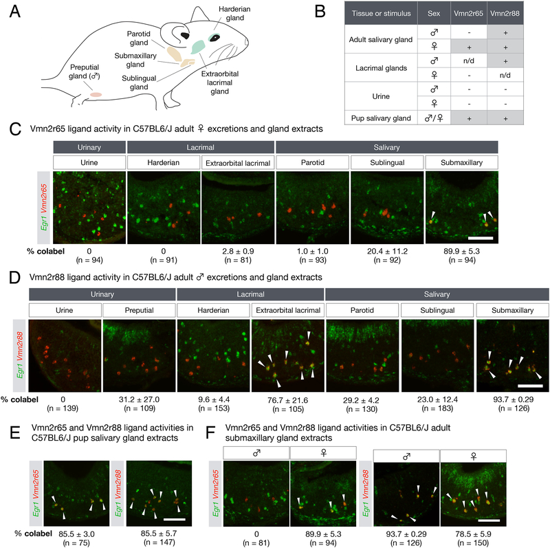
Figure 3: The origins of pup stimuli.
(A) Schematic diagram depicting the location of various exocrine glands (green: lacrimal, orange: salivary, pink: preputial) used to test Vmn2r65 and Vmn2r88 ligand activity.
(B) Summary of VR activity using gland extracts from adults and pups, and adult urine. n/d: not determined.
(C) Vmn2r65 ligand activity is found exclusively in female submaxillary gland extract. RNA FISH with Egr1 and Vmn2r65 probes on virgin male VNOs after exposure to urine and gland extracts from adult virgin females. Arrows mark Egr1 and Vmn2r65 co-expression.
(D) Vmn2r88 ligand activity is found in extracts of multiple glands. RNA FISH with Egr1 and Vmn2r88 probes on VNOs from virgin males after exposure to urine and gland extracts from adult virgin males. Arrows mark Egr1 and Vmn2r88 co-expression.
(E-F) Vmn2r88 and Vmn2r65 ligand activities are found in both pup and adult tissue extracts. All experiments were performed on 2 animals per stimulus, and the mean percentage of VR+Egr1+ neurons in VR+ cells in virgin male VNOs is indicated, along with the number of cells analyzed. All errors are in SEM.
All scale bars represent 100 μm.

