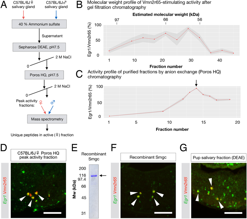
Figure 4: The submandibular gland protein C (Smgc) activates neurons expressing Vmn2r65.
(A) Purification strategy of compound(s) activating Vmn2r65 neurons (see Methods). The mass spectrometry profile of the peak activity fractions obtained from adult female salivary gland extracts was compared to the most homologous fractions obtained from male gland extracts.
(B) Activity profile of gel filtration chromatography fractions using female C57BL/6J salivary gland extract as a starting material. The ligand activity for Vmn2r65 was assayed by exposure of virgin males to selected fractions. Each dot represents the mean percentage of Vmn2r65+Egr1+ neurons among all Vmn2r65+ neurons from quantified on at least four VNO sections. Molecular weight for each fraction was estimated by SDS-PAGE. The ligand activity appears to elute broadly from this column, spanning 60 to 90 kDa.
(C) Activity profile of anion exchange chromatography fractions using Sepharose DEAE peak activity fractions as the input. The arrow marks the peak activity fraction (assayed in D), which was further analyzed by mass spectrometry.
(D) RNA FISH showing that Vmn2r65 neurons are activated by the exposure of virgin males to the peak fraction of anion exchange chromatography.
(E) SDS-PAGE gel of recombinant histidine-FLAG double-tagged Smgc produced in E. coli.
(F) Exposure of virgin males to recombinant Smgc results in specific activation of Vmn2r65+ VNO neurons. RNA FISH of VNO sections from virgin males exposed to recombinant Smgc shows co-localization of Egr1 and Vmn2r65 (arrows) (84.2 ± 5.0% of Vmn2r65+ neurons; Error in SEM; 166 Vmn2r65+ neurons from 3 animals examined).
(G) Fractionated C57BL6/J pup salivary gland extracts activate Vmn2r65-expressing neurons.
All scale bars represent 100 μm.

