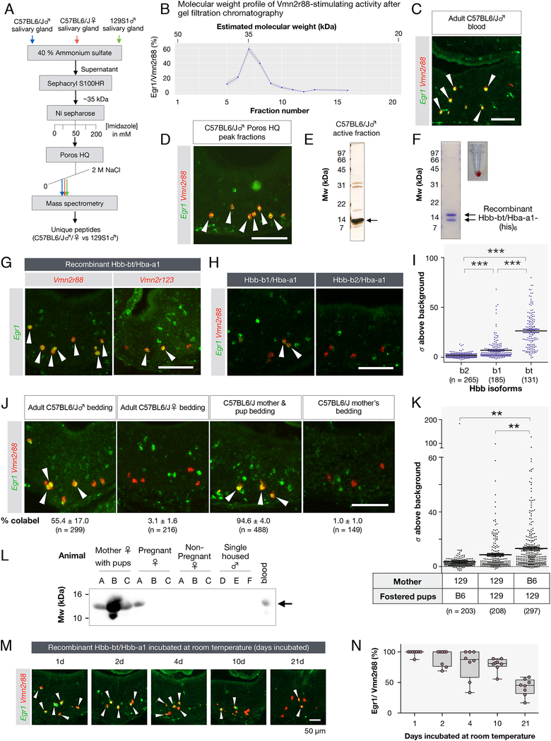
Figure 5: Hemoglobins are polymorphic ligands of Vmn2r88 and Vmn2r122/123.
(A) Purification scheme of the Vmn2r88 ligand (see Methods).
(B) Activity profile of C57BL6/J male salivary gland extracts after gel filtration chromatography. The activity was assessed as the percentage of Vmn2r88-expressing neurons with Egr1 signal.
(C) Adult C57BL/6J blood activates Vmn2r88-expressing VNO neurons (Male blood: 92.5 ± 1.8 % of Vmn2r88 neurons; Error in SEM; 161 Vmn2r88 neurons from 2 animals; Female blood: 97.3 ± 0.4 % of Vmn2r88 neurons; 152 Vmn2r88 neurons from 2 animals examined).
(D) Vmn2r88-stimulating activity of the purified Poros HQ peak fraction confirmed by RNA FISH. Arrows mark colocalization between Egr1 and Vmn2r88.
(E) Protein gel of the Poros HQ active peak fraction used for mass spectrometry.
(F) Recombinant hemoglobin produced in E. coli is red, indicating the presence of heme.
(G) Recombinant Hbb-bt/Hba-a1 robustly activates Vmn2r88-expressing neurons (94.1 ± 0.9 % of Vmn2r88 neurons; mean ± SEM; 240 Vmn2r88 neurons from 4 animals examined) and Vmn2r122/123-expressing neurons (63.4 ± 16.4 % of Vmn2r122/123 neurons; 184 Vmn2r122/123 neurons, 3 animals). Virgin males were exposed to recombinant Hbs and their VNOs were analyzed for the activation of Egr1 in Vmn2r88 or Vmn2r122/123 neurons. (H,I) Specificity of Vmn2r88 activation by Hbs. Virgin males were exposed to recombinant Hbb-b1/Hba-a1 and Hbb-b2/Hba-a1, and their VNOs were analyzed by RNA FISH with Egr1 and Vmn2r88 probes. The intensities of the Egr1 signals in Vmn2r88 neurons in Figure 5G,H, represented as fold increase from the standard deviation of image background, are plotted in a graph in Figure 5I. Each dot represents one cell. ***p < 0.001 by t-Test.
(J) Vmn2r88 ligand activity is deposited in bedding of males and in bedding of mothers cohabitating with infants. Mother/pup bedding robustly excites Vmn2r88 neurons, as evidenced by RNA FISH showing the colocalization of Egr1 and Vmn2r88 signals in the VNO of animals exposed to these cues (arrows). All experiments used 3 or 4 animals, and the percentage of Vmn2r88+Egr1+ cells in Vmn2r88+ neurons are indicated, along with the total number of cells analyzed. All errors are in SEM.
(K) Plot of Egr1 signals (represented as fold increase from the standard deviation of image background) in Vmn2r88 neurons, stimulated by bedding of cross-fostered cages. Bedding from cross-fostered cages (one 129S1/SvImJ mother with C57BL/6J pups, one 129S1/SvImJ mother with 129S1/SvImJ pups, one C57BL/6J mother with 129S1/SvImJ pups, N = 3 each). **p < 0.01 by Student’s t-Test.
(L) Hemoglobin is readily detectable in the cage of pregnant and post-partum females with pups. Shown is a protein blot of bedding extracts probed by anti-Hb beta antibody. Bedding from three females (A, B, C) for each condition and males (D,E,F) are used for this panel.
(M,N) Recombinant Hbb-bt/Hba-a1 was stored at room temperature for indicated period of time and tested for its ability to stimulate Vmn2r88-expressing neurons. Two CD-1 virgin males were used for this test per sample. The box plot in (N) shows the quantification of the Vmn2r88 neurons co-labeled with Egr1. 4 sections from 2 animals were quantified for each time point.
All scale bars represent 100 μm unless otherwise indicated.

