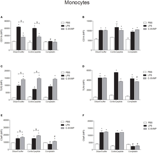
Figure 2.
Partially complement-dependent activation of monocytes by C-SVMP in the human whole blood model. Blood samples were collected in the presence of lepirudin and pre-incubated with diluent buffer, compstatin or control peptide for 30 min. Then, PBS, LPS or C-SVMP was added, and the samples were further incubated for 30 min. After erythrocyte lysis, leukocytes were fixed, stained with specific mouse anti-human monoclonal antibodies or isotype controls that were fluorochrome labeled and incubated for 30 min. Samples were analyzed by flow cytometry. Monocytes were gated based on their forward- and side-scatter features, followed by selection of the CD33+ population. The expression of CD11b (A), CD14 (B), TLR2 (C), TLR4 (D), C3aR (E), and C5aR1 (F) was determined by the median fluorescence intensity (MFI) of stained cells after subtraction of the respective isotype control. Data represent the mean ± S. D. of four independent experiments. *p < 0.05 compared to the respective PBS treatment. &p < 0.05 between the LPS and C-SVMP treatments. #p < 0.05 between the compstatin and diluent buffer pretreatments.

