
Figure 2.
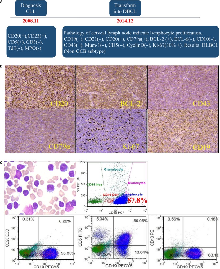
The transformation of the patient from CLL to DLBCL. A, Dynamic results pathology of lymph node during the progression of RS. B, Pathological assay of resected lymph node. C, BM tested by Pathology and immunophenotyping of this RS patient (87.8% cells were CD19 positive; CD5 partial positive, CD10 negative)